Take your research in any direction with the new benchmark for Raman imaging.
 Part of the Oxford Instruments Group
Expand
Collapse
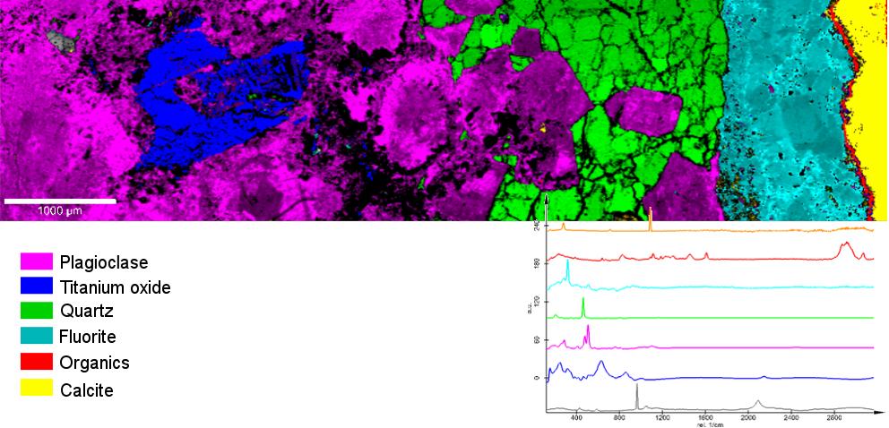
The Oxford Instruments Raman imaging systems are excellent analytical tools for the comprehensive investigation of geological samples, such as the identification and characterization of minerals, or in the observation of mineral phase transitions in high and ultra-high pressure/temperature experiments.
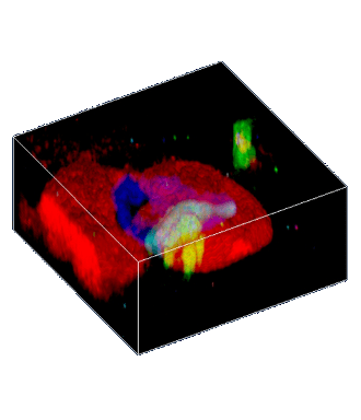
With confocal Raman imaging the spatial distribution and association of components or mineral phases, or chemical variation can be observed and this information may contribute significantly to the understanding of a sample’s complexity. Such characteristics can be evaluated with the witec360 microscope from large scale scans in the centimeter range to the finest detail with sub-micron resolution. Considering that most geo-materials are transparent from the NUV to VIS and NIR to some degree, this information can be furthermore obtained three-dimensionally due to the confocal set-up of the witec360 microscopes.
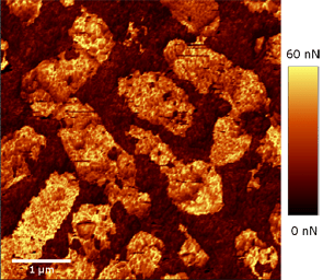
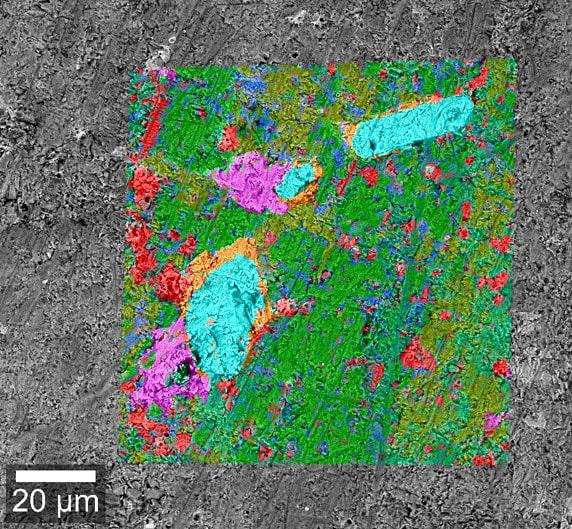
Oxford Instruments' highly versatile Raman microscopes can combine various imaging techniques to significantly increase the insights provided by measurement results. Possible combinations which can be included in a single microscope setup include confocal Raman imaging, Atomic Force Microscopy (AFM), Nearfield-Microscopy (SNOM) and Scanning Electron Microscopy (SEM). Confocal Raman imaging provides chemical information, AFM detects topography, structure, and physical properties such as stiffness, adhesion, etc. of the sample’s surface, and SNOM high-resolution measurements can optically reach beyond the diffraction limit. All witec360 configurations can be upgraded at any point to adapt the system to new or extended requirements.


If you'd like to learn more about the possibilities of Raman imaging for your individual field of application, one of our specialists will be happy to discuss them with you.
Contact us