Part of the Oxford Instruments Group
Expand

Atomic Force Microscopy (AFM)

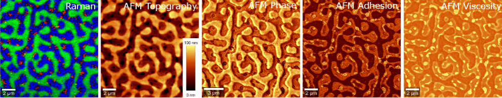
The AFM extension for witec360 Raman microscopes facilitates analysis of material surfaces at the nanometer scale. The integrated design allows for fast, seamless switching between AFM and other measurement modes, while also supporting simultaneous Raman-AFM measurements. With its combined approach, witec360 ensures precise optical alignment and enables convenient correlative imaging of the same sample area for comprehensive analyses in materials science, polymer research, nanotechnology, and life sciences.

You are interested in a stand-alone AFM?
Discover the Oxford Instruments Asylum AFM systems.


Request Pricing


Key Features

  • Easy combination of comprehensive surface characterization (AFM) and molecular information (Raman) in one system
  • Wide range of AFM modes for topographic, stiffness and adhesion investigations
  • High resolution capabilities: Lateral resolution down to 1nm; depth resolution <0.3 nm
  • Seamless combination with other correlative imaging techniques
  • Integrated measurement control and data analysis via the witec360’s Software Suite
  • Straight-forward navigation on the sample via high-quality video image
  • Easy exchange and alignment of premounted cantilevers
  • Precise sample positioning via TrueScan™ controlled piezo scan stages of selectable scan ranges
  • Microscope base with active vibration isolation system
  • Suitable for TERS applications
AFM Cat hair
AFM topography image of a cat hair.

AFM Operation Modes

  • Contact Mode
  • AC Mode (Intermittent Mode, Phase Imaging)
  • Digital Pulsed Force Mode (DPFM)
  • Lift Mode™
  • Magnetic Force Microscopy (MFM)
  • Electrostatic Force Microscopy (EFM)
  • Force Distance Curves
  • Lithography and Nanomanipulation
  • Lateral Force Microscopy (LFM)
  • Chemical Force Microscopy (CFM)
  • Kelvin Probe Microscopy
  • Conductive Contact AFM
  • Piezoresponse Force Microscopy
  • Operation in Liquid Environments
Raman-AFM of Polymers
Combined Raman-AFM measurement of the same sample area of a multicomponent polymer blend consisting of a 1:1:1 mixture of polystyrene, SBR (styrene-butadiene rubber) and EHA (2-ethylhexyl acrylate). Raman image (left): green = PS, red= SBR, blue = EHA.

The Principle

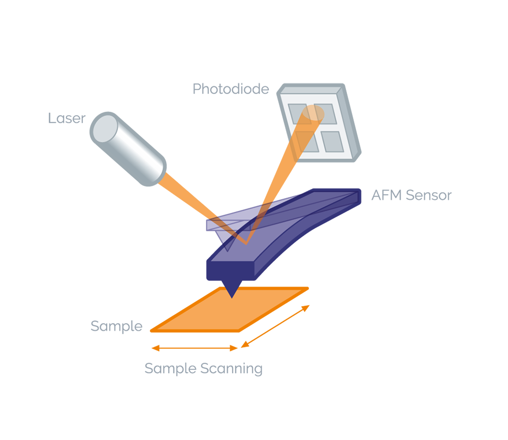
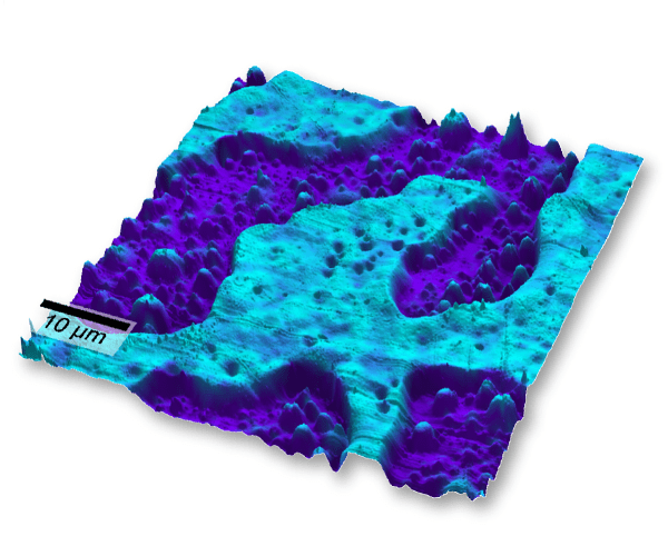
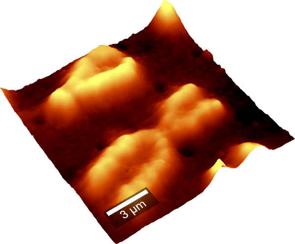
AFM measures surface topography and local material properties such as adhesion and stiffness by recording interaction forces between a sharp tip on a cantilever and the sample surface. The deflection of the cantilever is monitored as the sample is scanned with nanometer precision using a piezo-driven stage, enabling the generation of high-resolution images.

AFM Principle

Application Examples

Raman AFM stress in silicon
Correlative Raman-AFM microscopy of stress in silicon. Left: 10 μm x 10 μm AFM topography image and depth profile (bottom). Right: Corresponding Raman image revealing the stress areas in silicon. Sampe courtesy: Helmut Fischer GmbH, Sindelfingen, Germany.
Raman AFM polymer blend
Overlay of Raman and AFM image of a polymer blend of PS and PMMA.
AFM image of a human chromosome.

Literature

Application Note Correlative Raman Imaging in Polymer Research

Technical Note Correlative Raman AFM


Contact

Please fill in all data fields to ensure we can process your inquiry as quickly as possible.


Related Applications

Coatings & Thin FilmsGeo ScienceLife ScienceNano-Carbon & 2D MaterialsSemiconductors & Photovoltaics