 Part of the Oxford Instruments Group
Expand
Collapse
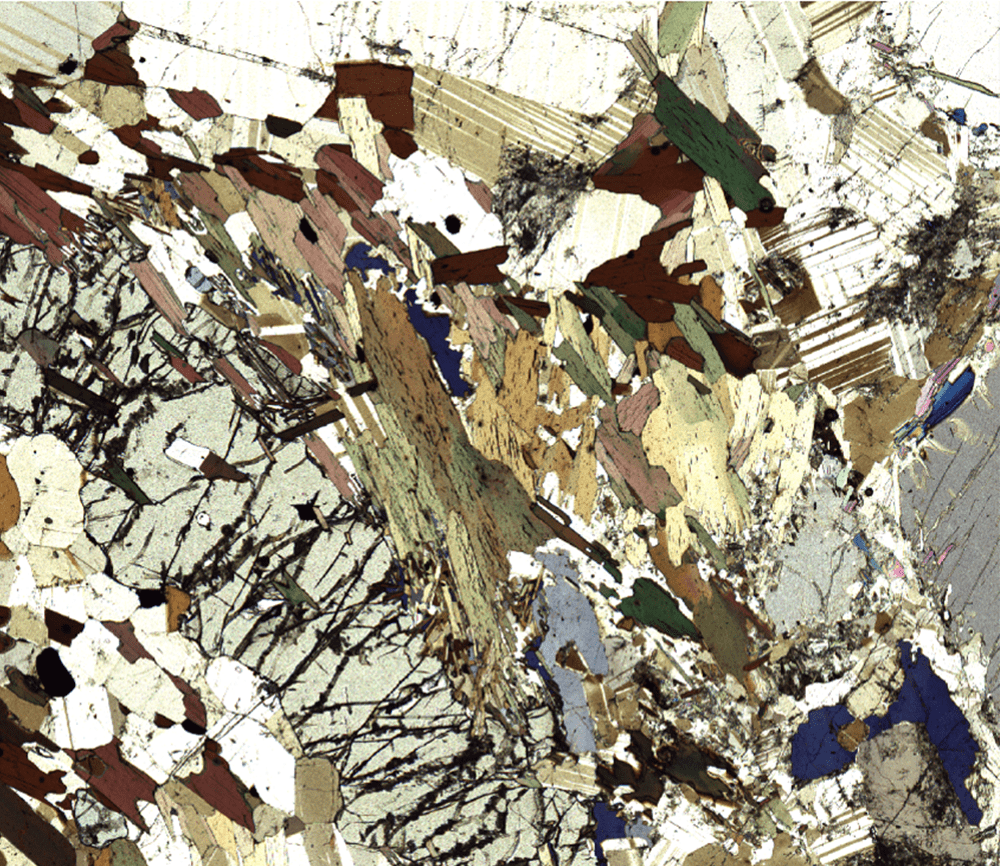
Oxford Instruments witec360 imaging microscopes are equipped with a Köhler-illumination setup with optimized optical components for generating white-light images of outstanding quality. We offer different options including bright- and dark-field imaging and differential interference contrast (DIC). White-light microscopy is possible in reflection and transmission modes and can feature polarization setups.
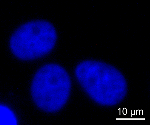
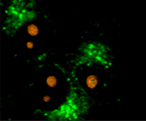
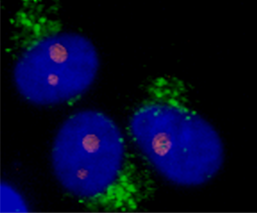
Optionally, the witec360 systems can integrate fluorescence microscopy for correlative approaches.
The witec360 imaging microscopes can integrate bright-field options in reflection or transmission mode, in addition to other techniques including differential interference contrast (DIC) and dark-field imaging.
All light microscopy techniques in witec360 systems feature software-controlled focus stacking and image stitching. With focus stacking, sharp images of samples of varying height can be quickly generated from a stack of individual images. Automated image stitching combines a series of individual measurements into a large-area image at the highest resolution.
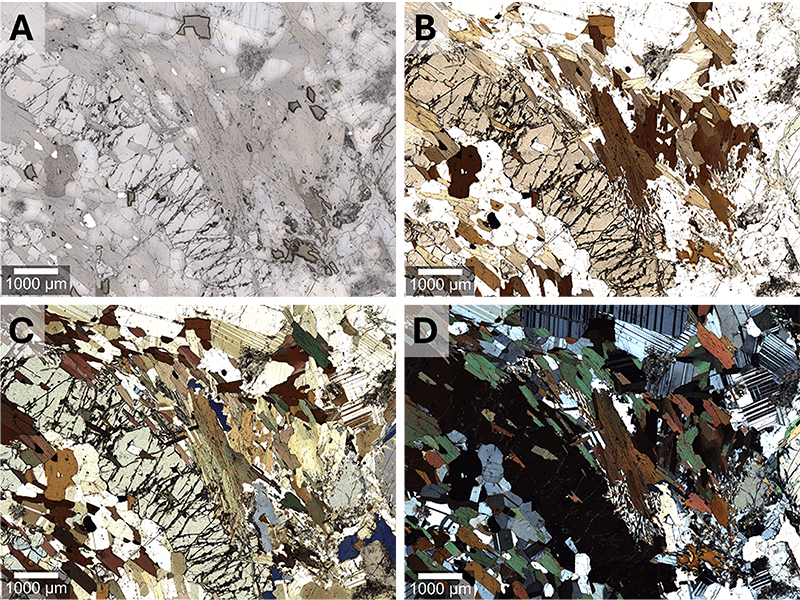
White-light microscopy in witec360 microscopes can optionally be upgraded with polarization modules, ideally suited for applications in geology, biology, chemistry and material science. The independent control of the polarizer (excitation path) and analyzer (detection path) facilitate different polarization illumination setups in parallel and perpendicular orientations.
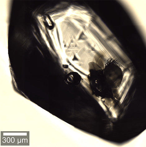
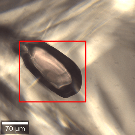
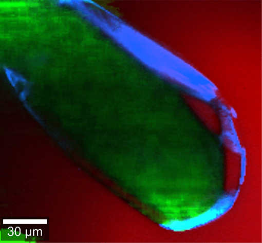
The white-light microscopy capabilities of the witec360 systems convince with their exceptional performance. In this example, small inclusions were easily found in a diamond, even though they were located 20 – 50 µm beneath the surface. Confocal Raman imaging at the same position then revealed the inclusions' composition.
Combine Raman microscopy and fluorescence imaging in one instrument. The optional fluorescence setup for witec360 microscopes uses laser excitation and features a highly sensitive monochromatic camera for detection. Matching emission filters are available for a large number of compatible wavelength combinations.
Please fill in all data fields to ensure we can process your inquiry as quickly as possible.