witec360 Raman microscope with Hexalight spectrometer launched
Take your research in any direction with the new benchmark for Raman imaging.
 Part of the Oxford Instruments Group

The non-invasive and label-free nature of confocal Raman microscopy makes it a powerful technique for cell biology applications. By using laser excitation and the inelastic scattering of light by molecules, it provides valuable chemical and structural information about the cellular environment. As a result, you can visualize different biomolecules, including lipids, proteins and nucleic acids and their distribution in the cell. Current research in cell biology uses Raman imaging particularly to study cellular processes such as lipid organization, metabolism, infection and drug uptake [1-7]. The high spatial resolution and chemical specificity of confocal Raman imaging make it a valuable tool for understanding the complex dynamics of cells in various biological processes.
The outstanding technology in Oxford Instruments' Raman systems provides highest resolution, speed and sensitivity simultaneously, to deliver the best results from your valuable samples. With their unmatched versatility, the witec360 Raman microscopes are the optimal base to adapt to your research requirements now and in the future. For biological applications that work with living specimens, we offer compatible devices for environmental and temperature control.
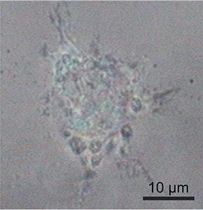
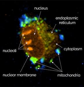
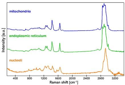
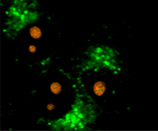
Since confocal Raman imaging is a non-destructive microscopy technique, it is well suited for studying living cells in their physiological surroundings without damaging them. In this example, epithelial rat cells were investigated. Typical Raman spectra were found for mitochondria (blue), endoplasmic reticulum (green) and nucleoli (orange). The color-coded Raman image visualizes the location of these organelles in the cell.
Read more in our Application Note
The combination of confocality and excellent depth resolution in Oxford Instrument witec360 systems enables you to resolve the details of cells even in 3D. This animation shows a 3D Raman image of a mesenchymal cell in hydrogel and visualizes the different cellular components that were identified 1.
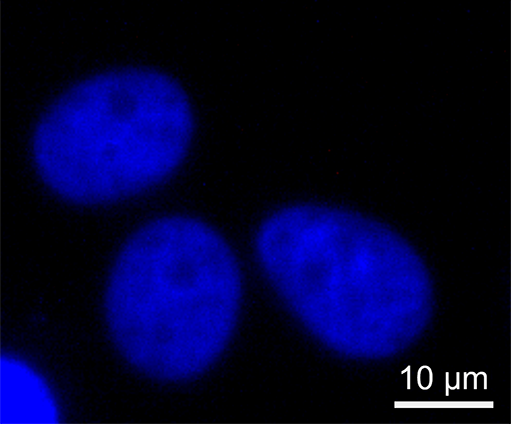
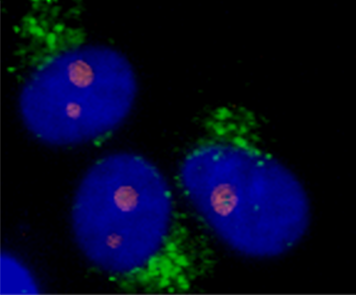
In cell biology research, fluorescent labeling is a commonly used method to visualize specific molecules. Although Raman imaging provides a powerful alternative, both techniques can optionally be combined in witec360 microscopes for those applications that require additional fluorescent markers. These pictures show the correlative Raman and fluorescence imaging of DAPI-stained eukaryotic cells. The fluorescence image visualizes the labeled cellular nuclei, while Raman imaging identified the endoplasmic reticulum (green) and nucleoli (orange).
Read more in our Application Note
Oxford Instruments' highly versatile Raman microscopes can combine Raman with various other imaging techniques to provide complementary information from the same sample position.


Application Note Confocal Raman Imaging and Correlative Techniques in Life Science
Customer publications:
If you'd like to learn more about the possibilities of Raman imaging for cell biology applications, one of our specialists will be happy to discuss them with you.
Contact us
© Oxford Instruments 2026