 Part of the Oxford Instruments Group
Expand
Collapse
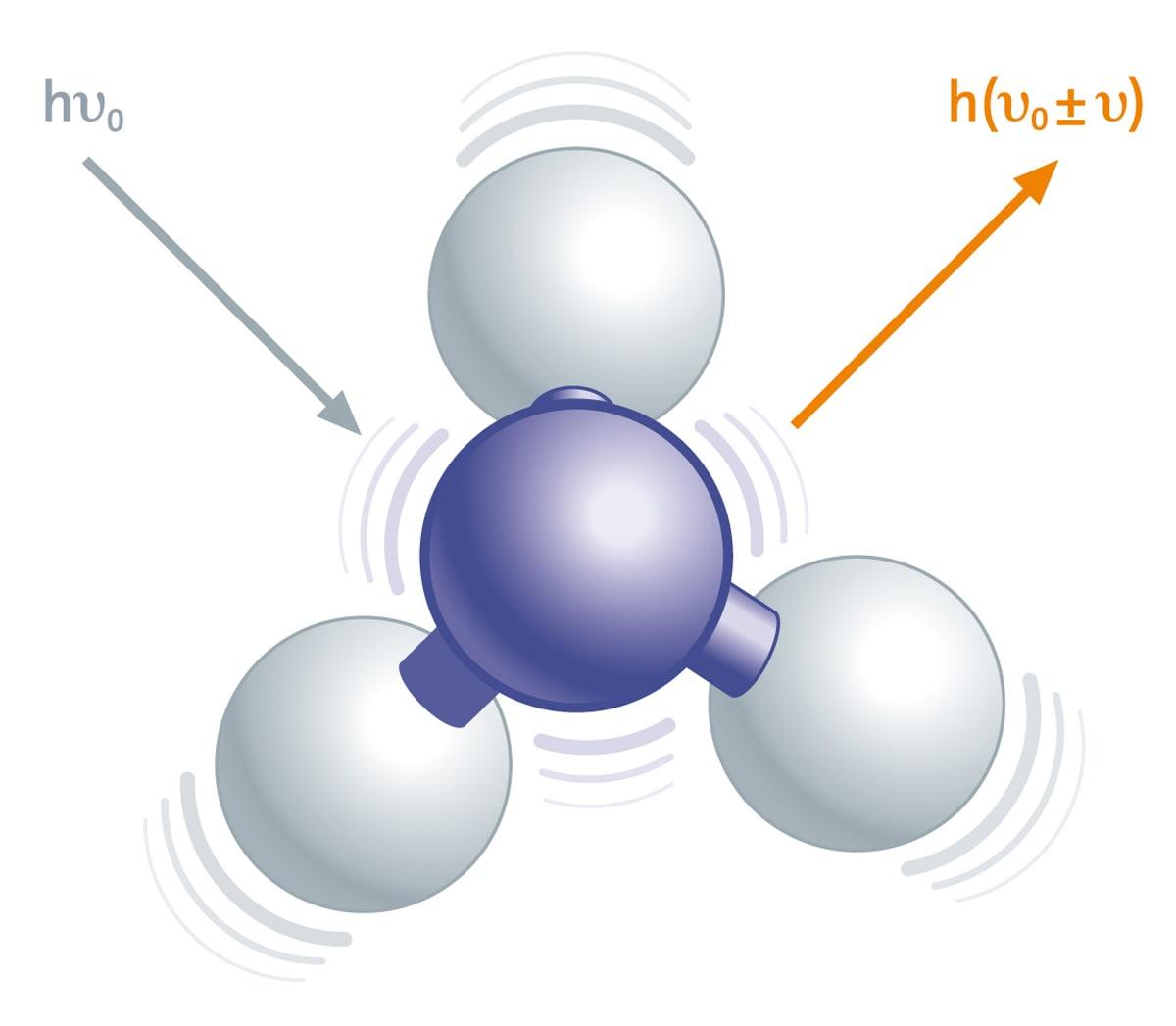
The Raman effect is based on light interacting with the chemical bonds of a sample. Due to vibrations in the chemical bonds the interaction with photons causes specific energy shifts in the back scattered light that appear in a Raman spectrum. The Raman spectrum is unique for each chemical composition and can provide qualitative and quantitative information of the material.
Confocal Raman microscopy is a high-resolution imaging technique that is widely used for the characterization of materials and specimens in terms of their chemical composition. Chemical properties of solid and liquid components can be analyzed with diffraction-limited spatial resolution (λ/2 of the excitation wavelength, down to 200 nm). No labeling or other sample preparation techniques are necessary. With Raman images, information regarding the chemical compounds and their distribution within the sample can be illustrated clearly.
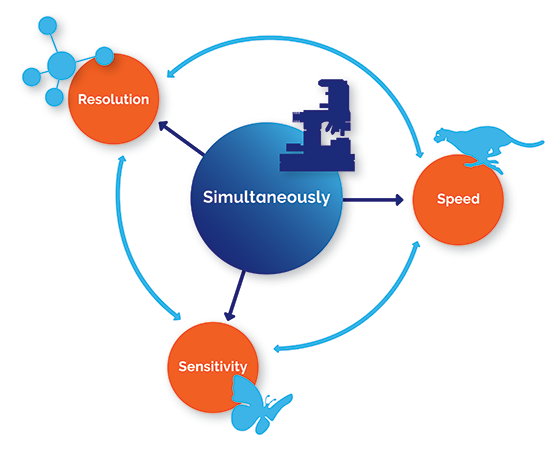
Oxford Instruments' Raman microscopes and imaging systems combine an extremely sensitive confocal microscope with an ultra-high throughput spectroscopy system for unprecedented chemical sensitivity. Their outstanding performance in speed, sensitivity and resolution can be jointly applied without compromises.
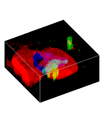
3D volume scans and depth profiles are valuable tools in providing information about the dimensions of objects or the distribution of a certain compound throughout the sample.
Oxford Instruments' confocal microscope systems provide depth resolution and a strongly reduced background signal and facilitate the generation of depth profiles and 3D images with exceptional spectral and spatial resolution. Images are recorded point by point and line by line, while scanning the sample through the excitation focus. With this technique, the specimen can be analyzed in segments along the optical axis and depth profiles or 3D images can be generated.
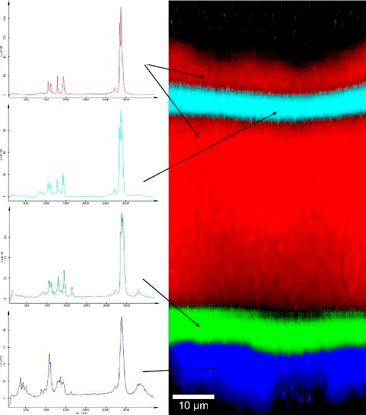
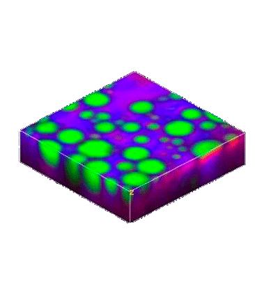
With ultra-fast Raman imaging it is possible to acquire a complete Raman image in a few minutes. In other words the acquisition time for a single Raman spectrum can be as low as 760 microseconds, 1300 Raman spectra can be acquired per second.
The latest spectroscopic EMCCD detector technology combined with the high-throughput optics of a confocal Raman imaging system are the keys to this improvement which can also be advantageous when performing measurements on delicate and precious samples requiring the lowest possible levels of excitation power. Time-resolved investigations of fast dynamic processes can also benefit from the ultra-fast spectral acquisition times.

Please fill in all data fields to ensure we can process your inquiry as quickly as possible.