Take your research in any direction with the new benchmark for Raman imaging.
 Part of the Oxford Instruments Group
Expand
Collapse
The development and production of drug delivery systems requires efficient and reliable control mechanisms to ensure the quality of the final products. These products can vary widely in composition and application. Therefore analytical tools such as the Oxford Instruments witec360 microscopes that provide both comprehensive chemical characterization and the flexibility to adjust the method to the investigated specimen are preferred in pharmaceutical research.
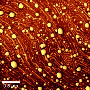
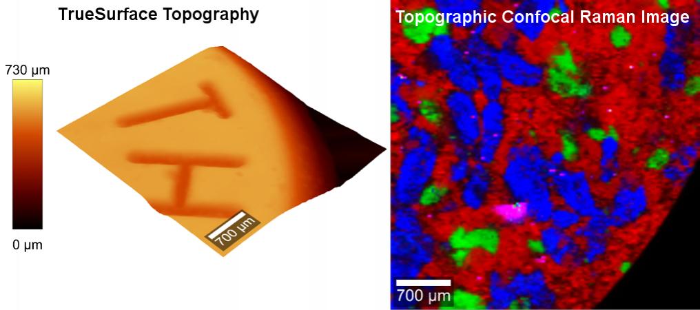
Oxford Instruments' highly versatile Raman microscopes can combine various imaging techniques to significantly increase the insights provided by measurement results. Possible combinations which can be included in a single microscope setup include confocal Raman imaging, Atomic Force Microscopy (AFM), Nearfield-Microscopy (SNOM) and Scanning Electron Microscopy (SEM). Confocal Raman imaging provides chemical information, AFM detects topography, structure, and physical properties such as stiffness, adhesion, etc. of the sample’s surface, and SNOM high-resolution measurements can optically reach beyond the diffraction limit. All witec360 configurations can be upgraded at any point to adapt the system to new or extended requirements.
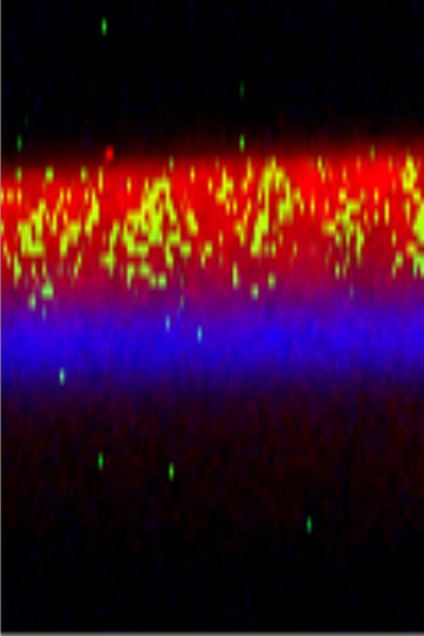
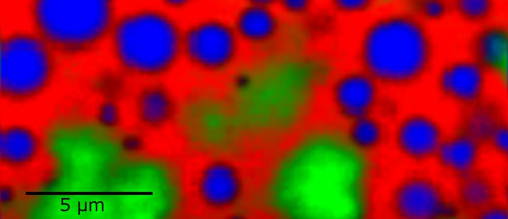
Confocal Raman imaging can be used to survey the distribution of components within formulations, to characterize homogeneity of pharmaceutical samples, to determine the solid state of drug substances and excipients and to characterize contaminations and foreign particulates. The information obtained by confocal Raman microscopy is also extremely useful for drug substance design, for the development of solid and liquid formulations, as a tool for process analytics and for patent infringement and counterfeit analysis.


If you'd like to learn more about the possibilities of Raman imaging for your individual field of application, one of our specialists will be happy to discuss them with you.
Contact us