 Part of the Oxford Instruments Group
Expand
Collapse

StrobeLock is an extension for Oxford Instruments' witec360 microscopes, which enables time-correlated single photon counting (TCSPC) measurements. TCSPC records the time-resolved luminescence or fluorescence decay after pulsed stimulation. In Time-resolved Luminescence Microscopy (TLM), the intensity decay is recorded at each image pixel. Various parameters describing each decay can be determined, such as single- or even multi-exponential relaxation times. Images can then be color-coded according to any of the extracted parameters, revealing local differences in the emission behavior of a sample. For example, extracting the lifetime of fluorophores allows for Fluorescence Lifetime Imaging (FLIM). Combining the time-resolved data with Raman, AFM or SNOM images yields additional information on the investigated materials.
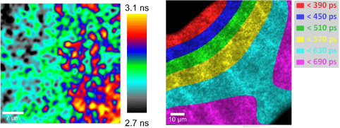
With Time-resolved Luminescence Microscopy (TLM), the luminescence properties of light-emitting devices, such as LEDs, can be investigated and spatial differences can be revealed. Light emission is stimulated by an electrical pulse generator and its intensity is recorded in a time-resolved manner. Thus, relaxation times or response times can be calculated for each image pixel and visualized. TLM can be used for example for the quality control of LEDs.

Time-resolved Luminescence Microscopy (TLM) on a blue light-emitting diode (LED).
Left: Map of local relaxation times; scale bar 7 µm.
Right: Contour plot of the temporal start of the luminescence emission; scale bar 10 µm.
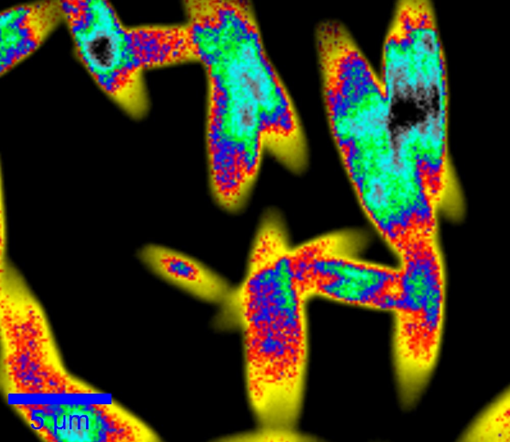
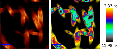
Fluorescence Lifetime Imaging Microscopy (FLIM) determines the average fluorescence lifetime for each image pixel from the time-resolved fluorescence decay after excitation by a pulsed laser. The resulting FLIM image is color-coded according to the lifetime, displaying its spatial distribution in a sample. In combination with other imaging techniques, such as Raman imaging or AFM, FLIM extends the amount of information gained from one sample.

Fluorescence Lifetime Imaging (FLIM) of N,N′‐bis(1‐ethylpropyl)‐3,4,9,10‐perylenebis(dicarboximide) (EPPTC) crystal needles. Left: Total fluorescence intensity; scale bar 5 µm. Right: FLIM; scale bar 5 µm.
Images are a courtesy of Xinping Zhang, Institute of Information Photonics Technology and College of Applied Sciences, Beijing University of Technology.
Please fill in all data fields to ensure we can process your inquiry as quickly as possible.