Part of the Oxford Instruments Group
Expand
Collapse
Part of the Oxford Instruments Group

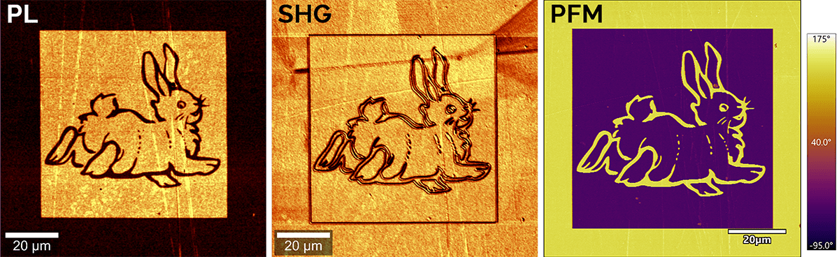
Oxford Instruments' witec360 Raman microscopes can seamlessly integrate nonlinear optical imaging techniques such as second-harmonic generation (SHG) and third-harmonic generation (THG) imaging, to provide advanced solutions for comprehensive material characterization. This combined approach eliminates the need for separate costly equipment and lowers the barriers to adopting these methods, which are particularly valuable for 2D materials research, ferroelectric material development and bio imaging.
The witec360 supports SHG and THG measurements under extreme conditions, including low/high temperatures, high pressure, and magnetic fields. For precise analysis of crystalline orientation and chirality using SHG, the microscope enables polarization-resolved measurements with unlimited flexibility in polarization direction.
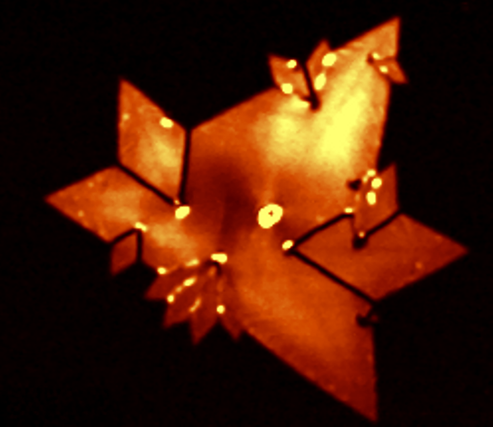
Second Harmonic Generation (SHG) is a nonlinear optical process that converts two photons of incident light into a single photon of emission. SHG depends on the non-centrosymmetric symmetry of a material’s structure and is highly responsive to variations in crystal orientation, crystal symmetry, layer thickness, and stacking order.
The physical properties of 2D materials, including transition metal dichalcogenides (MX₂) and hexagonal boron nitride (hBN), are strongly influenced by factors such as crystal symmetry, thickness, and stacking order. Combined with atomic force microscopy (AFM) and piezoresponse force microscopy (PFM), SHG is a valuable tool for analyzing the properties of piezoelectric and ferroelectric materials.
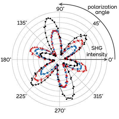
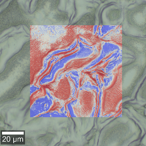
Polarization-resolved SHG measurements provide detailed insights into the crystal orientation in 2D materials, enabling precise structural characterization and analysis of grain alignment.
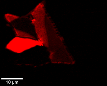
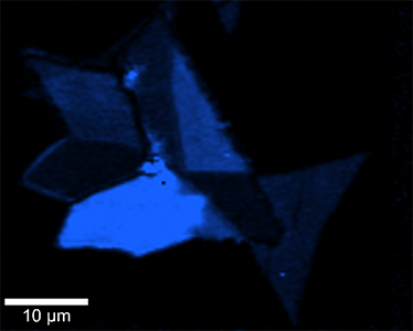
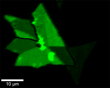
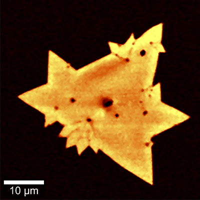
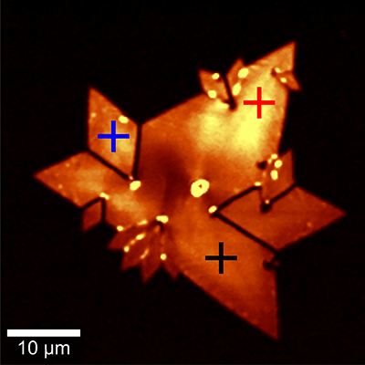
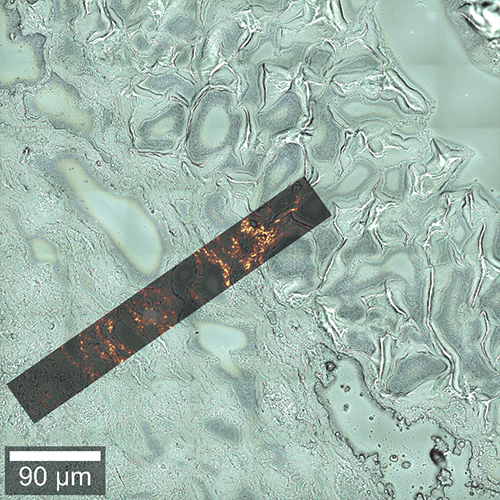
SHG imaging is highly sensitive to grain boundaries in 2D materials, as demonstrated on this example of an MoS₂ flake. Polarization-resolved SHG revealed differences in crystal orientation between grains and detected strain fields, based on the asymmetric patterns in the polar plots at the different positions in the flake.
Read more about the correlative analysis of MoS₂ using Raman, SHG and PL in our Application Note.
Nonlinear optical microscopy for bio-imaging provides deep insights into tissue morphology, structure, and cellular organization. It is commonly applied in cell biology, developmental biology, and disease research.
SHG imaging visualizes non-centrosymmetric structures, such as collagen in the extracellular matrix (ECM) and myosin filaments, offering information on tissue remodelling, cell migration, and fibrosis. THG imaging detects structural interfaces, enabling label-free imaging of cellular and subcellular features, including membranes, organelles, and lipid droplets, in their natural state.
Application Note
Correlative Imaging of 2D Material Heterostructures: Raman, PL and SHG
Application Note
Correlative High-resolution Imaging of TMDs
Application Note
Confocal Raman Imaging and Correlative Techniques in Life Science
Please fill in all data fields to ensure we can process your inquiry as quickly as possible.

© Oxford Instruments 2026