Part of the Oxford Instruments Group
Expand
Collapse
Part of the Oxford Instruments Group

Oxford Instruments' witec360 spectroscopy and microscopy systems can be configured with our flexible and automatable polarization modules for advanced structural and molecular analysis approaches. We offer solutions for polarization-resolved measurements in a variety of confocal optical techniques, such as white light imaging and spectroscopic applications.
The freely rotatable linear polarizer and analyzer modules enable you to perform spectroscopic measurements at any desired polarization configuration without sample rotation and/or adjustment of the sample position. Additionally, motorized modules allow for comfortable and precise serial recordings at fixed or flexible angular offsets between the polarizer and analyzer.
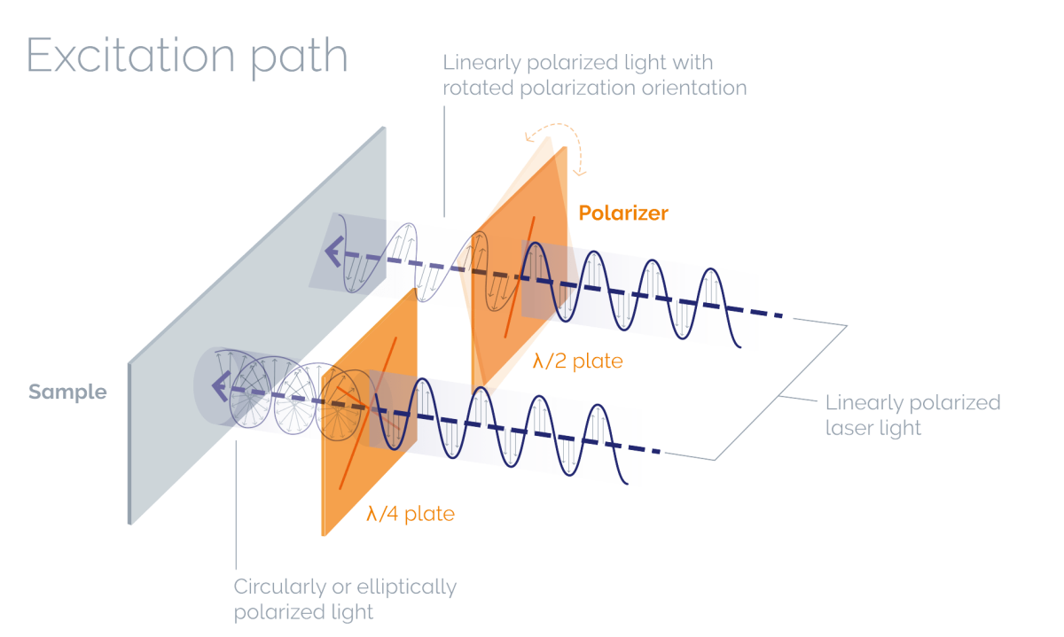
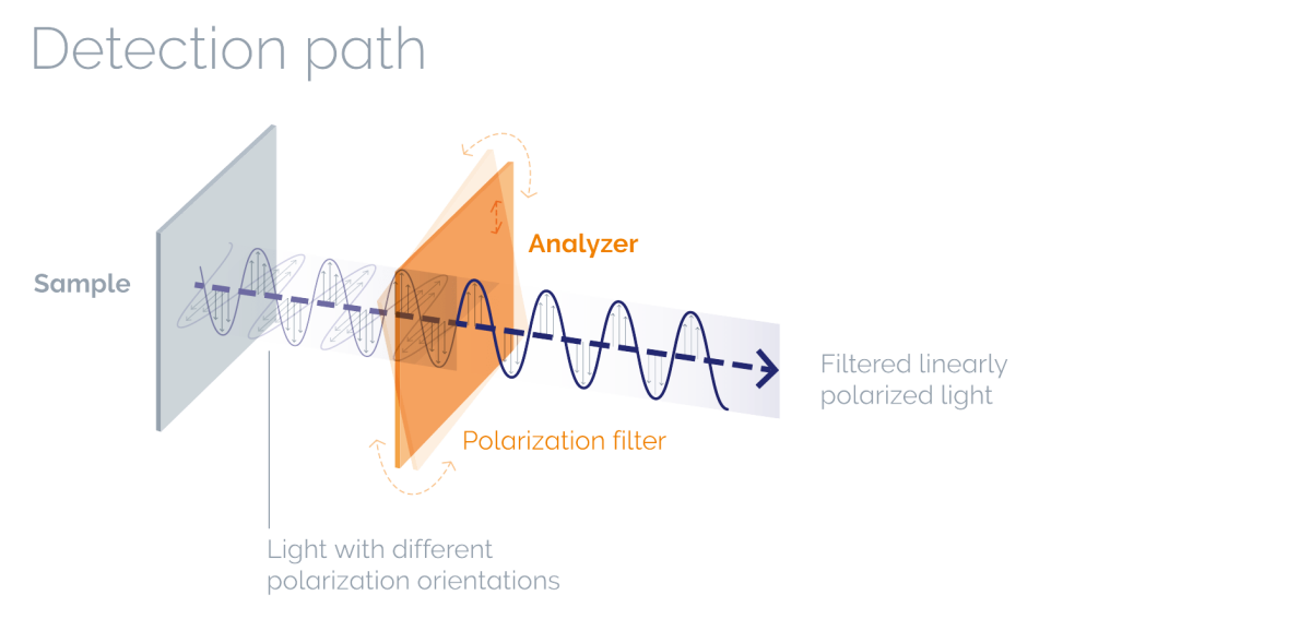
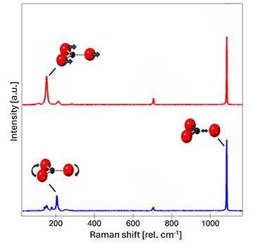
Waveplates in the polarizer module can rotate the linearly polarized light from the laser in a defined direction (half wave plate, λ/2) or transform it into circularly or elliptically polarized light (quarter wave plates, λ/4). A polarization filter in the analyzer within the detection path filters the light from the sample along a selectable polarization axis before detection.
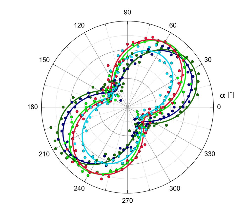
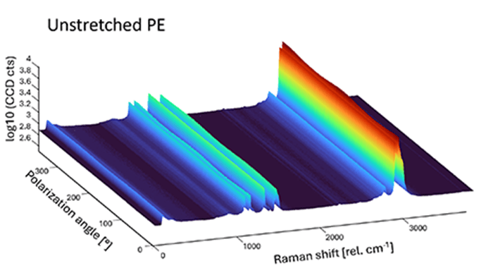
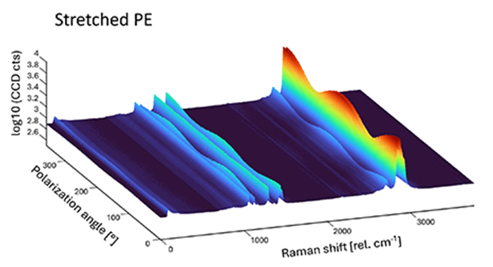
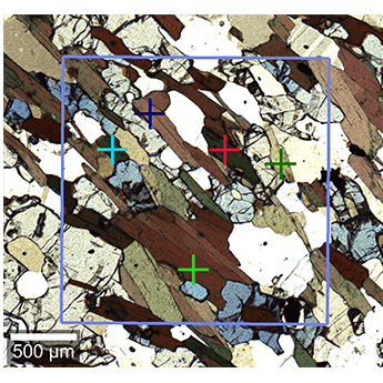
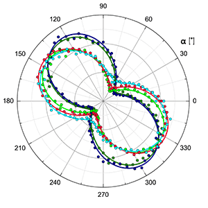
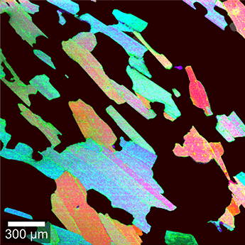
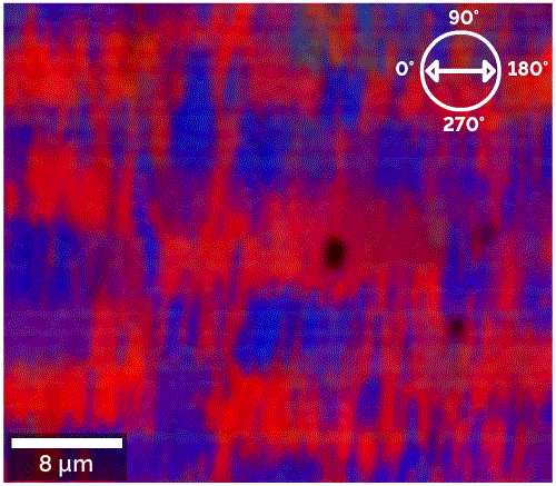
Using polarization analysis in combination with Raman spectroscopy enables the probing of advanced molecular information of the sample including the symmetry of the respective vibrations. As such, the orientation of different domains, chirality and optical anisotropy can easily be visualized.
Technical Note Polarization-resolved Raman microscopy and spectroscopy
Please fill in all data fields to ensure we can process your inquiry as quickly as possible.

© Oxford Instruments 2026