Part of the Oxford Instruments Group
Expand

RISE – Raman-SEM Microscopes

World's first fully integrated Raman Imaging and Scanning Electron Microscopes

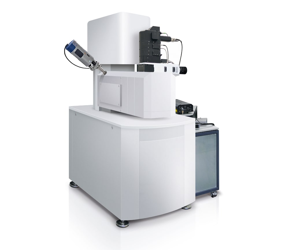
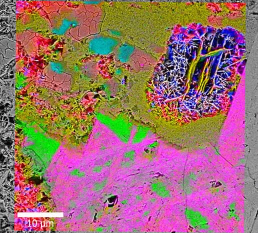
RISE Microscopy is a novel correlative microscopy technique that combines SEM and confocal Raman Imaging. Through RISE Microscopy ultra-structural surface properties can be linked to molecular compound information.

The RISE Microscope combines all features of a stand-alone SEM and the confocal Raman imaging microscope alpha300 within one instrument:

  • Quick and convenient switching between Raman and SEM measurement
  • Automated sample transfer from one measuring position to the other
  • Integrated software interface for user-friendly measurement control
  • Correlation of the measurement results and image overlay
  • No compromise in SEM and Raman imaging capabilities


Request Pricing


Literature

WITec RISE Brochure

RISE/EDS Brochure

Application Note RISE Microscopy (Raman-SEM)

Application Note Raman-SEM (RISE) Imaging of 2D Materials


The RISE Technique

Technique RISE

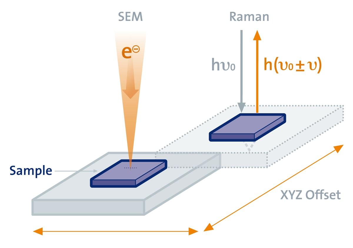
For RISE microscopy samples are automatically transferred from one measuring position to the other within the vacuum chamber of the SEM, streamlining the workflow and drastically improving the instrument's ease of use.


Application Examples

RISE Raman SEM Brain Tissue
Raman-SEM image overlay of a hamster brain tissue sample. Color-coded Raman image parameters: Green: White brain matter; Red: Gray brain matter; 100 µm x 100 µm, 300 x 300 pixels = 90,000 spectra, 50 ms integration time per spectrum.
RISE Raman SEM GaAs
Raman-SEM image overlay of a GaAs semiconductor sample. Color-coded Raman image parameters: Yellow: Gold substrate; Red: GaAs; Blue: Residues from production; 50 µm x 50 µm, 300 x 300 pixels = 90,000 spectra, 34 ms integartion time per spectrum.
RISE Raman SEM Polymers
Raman-SEM image overlay of a PMMA-PS polymer blend- Color-coded Raman image parameters: Green: Polystyrene; Red: Polymethyl methacrylate.
RISE Raman SEM Diorite
RISE Microscopy and EDX analysis of a geological sample. Left: Overlaid SEM-EDX image: Three different element groups can be distinguished (Orange: Si, O; Purple: Si, Al, Fe, Ca; Green: Na). Middle: The Raman-SEM image overlay of the same sample area shows the distribution of the molecular compounds. Right: Corresponding Raman spectra.

RISE Key Features

Scanning Electron Microscopy:

Scanning electron microscopy
Scanning Electron Microscopy features a variety of analysis techniques. With RISE, the chemistry of molecular compounds can be imaged additionally.

Confocal Raman Microscopy:

  • Confocal Raman Imaging with unprecedented performance in speed, sensitivity, and resolution
  • Hyperspectral image generation with the information of a complete Raman spectrum at every image pixel
  • Excellent diffraction limited lateral resolution of 200 - 300 nm
  • Outstanding depth resolution ideally suited for 3D image generation and depth profiles
  • Ultrahigh-throughput lens-based spectroscopic system for highest sensitivity and best performance in spectral resolution
  • Ultra-fast Raman imaging option with only 0.76 ms integration time per spectrum
  • Non-destructive imaging technique: no staining or fixation of the sample required

RISE Microscopy is suitable for you...

...as a Raman newcomer you will benefit from the ease-of-use and intuitive measurement procedure

...as an experienced user you will appreciate the exceptional correlative microscope performance with the advantages of both techniques included in one instrument

Materials sciene, nanotechnology, polymer science, geoscience, life science, pharmaceutical industry...

...regardless of the field in which you are working, RISE Microscopy will empower you with its unique imaging capabilities


Award Winning Technology

RISE Microscopy has been recognized with a 2015 Photonics Prism Award. An expert jury named the correlative RISE microscope as winner in the metrology category. The Prism Award is given for top innovations in the field of photonics, granted by Photonics Media and sponsored by the international Society for Optics and Photonics (SPIE).

DrkGreenWinnerBox

Contact

Please fill in all data fields to ensure we can process your inquiry as quickly as possible.


Related Applications

Food TechnologyForensicsArchaeology & The ArtsCoatings & Thin FilmsEnergy StorageGeo ScienceLife ScienceMaterials ScienceNano-Carbon & 2D MaterialsPharmaceutics & CosmeticsPolymersSemiconductors & Photovoltaics