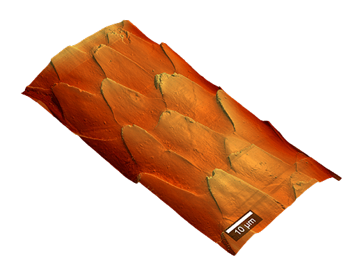
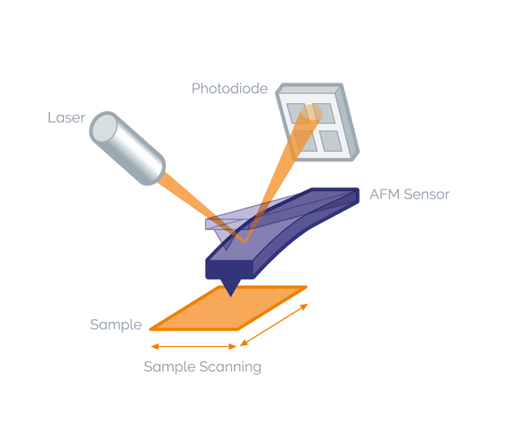
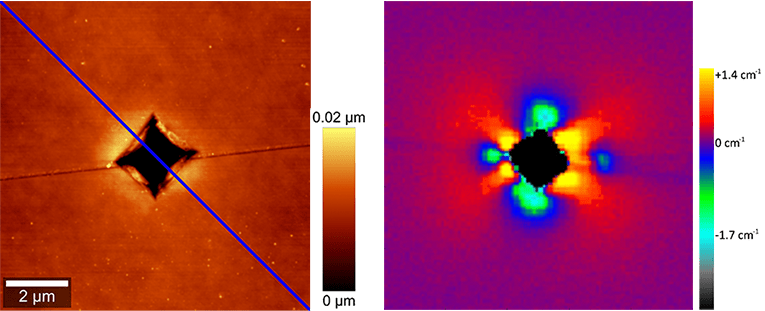
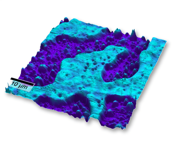
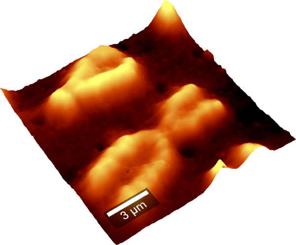
Atomic Force Microscopy (AFM) traces the topography of samples with extremely high - up to atomic - resolution by recording the interaction forces between the surface and a sharp tip mounted on a cantilever. AFM provides spatial information parallel and perpendicular to the surface. In addition to topographic high-resolution information, local material properties such as adhesion and stiffness can be investigated by analyzing tip-sample interaction forces.
The WITec Atomic Force Microscope integrated into a research-grade optical microscope provides superior optical access, exact cantilever alignment and high-resolution sample survey. The sample is scanned under the tip using a piezo-driven scanning-stage and the results (e.g. topography etc.) are displayed as an image.
The optical mode combined with an advanced video camera system allows high-resolution sample survey and quick selection of the area-of-interest. Through the simultaneous sample and AFM-cantilever view, the measurement position can be easily identified and adjusted. Using additional illumination and detection techniques (e.g. bright-field, dark-field, polarization, fluorescence, etc.), the user can further determine the point-of-interest for the AFM measurement. By simply rotating the microscope turret, the user can switch between optical and AFM modes quickly and accurately, maintaining the look and feel of optical microscopy in the AFM mode.
The Digital Pulsed Force Mode (DPFM) is a non-resonant, intermittent-contact mode. It avoids the surface damage that can result from operating in contact mode on soft samples and provides additional sample information.
When added to an AFM system, the DPFM extends the capabilities of the microscope beyond simply measuring topography to properties such as local stiffness, adhesion, viscosity, energy dissipation, contact time, long range forces etc. This can be done at normal scan rates, because the system works at up to several thousand pixels per second.
The electronics of the DPFM include a high speed data-acquisition system, a freely programmable modulation generator and a real-time data evaluation module. With the storage of the complete measurement, extensive post-processing data evaluation is performed.
The modular design of the WITec systems allows to combine various imaging techniques such as Raman imaging, fluorescence, luminescence, atomic force microscopy (AFM) and near-field microscopy (SNOM or NSOM) and in one single instrument for a more comprehensive, correlative sample analysis. Switching between the different modes is simply done by rotating the microscope turret.
By coupling confocal Raman imaging with AFM, the chemical properties of the sample can be easily correlated with its surface structure. These two complementary techniques are available in our combinatorial Raman-AFM microscope alpha300 RA for flexible and comprehensive sample characterization.
TERS (Tip-Enhanced Raman Spectroscopy) is a typical application for correlative Raman-AFM microscopes.
Please fill in all data fields to ensure we can process your inquiry as quickly as possible.