Part of the Oxford Instruments Group
Expand

Scanning Near-field Optical Microscopy (SNOM)

WITec SNOM Principle

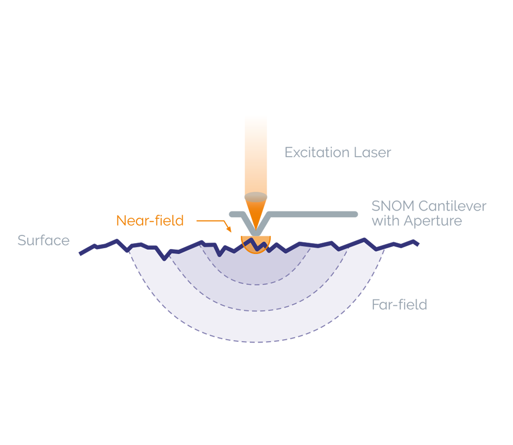
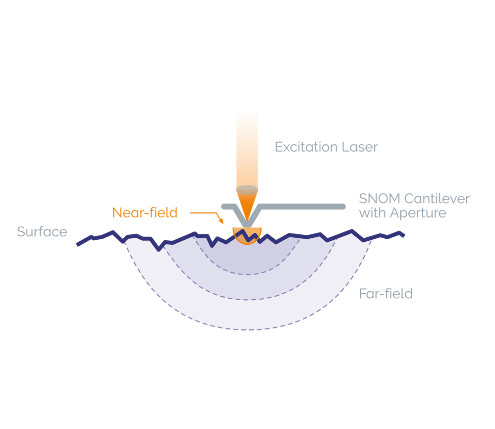
In Scanning Near-field Optical Microscopy, the excitation laser light is focused through an aperture with a diameter smaller than the excitation wavelength, resulting in an evanescent field (or near-field) on the far side of the aperture. When the sample is scanned at a small distance below the aperture, the optical resolution of transmitted or reflected light is limited only by the diameter of the aperture. The optical resolution attainable is in the range of 60 – 100 nm. The optical image is generated by scanning the sample's surface point-by-point and line-by-line.

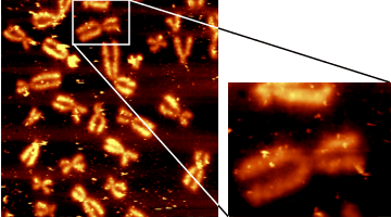
Typical applications are found in nanotechnology research and in particular the highly relevant fields of nano-photonics and nano-optics. In life science and materials research, SNOM allows the optical detection of the most miniscule surface structures of transparent as well as opaque samples. Using combinations with fluorescence techniques, even single-molecule detection can be easily achieved.


WITec’s Scanning Near-field Optical Microscope with Unique Cantilever Technique

With featured SNOM objectives and unique cantilever SNOM sensors, imaging beyond the diffraction limit is accomplished quickly and effortlessly with the WITec SNOM microscopes:

All WITec SNOM microscopes are equipped with unique, patented, high-quality micro-fabricated SNOM sensors, consisting of a silicon cantilever with a hollow aluminum pyramid as a tip. The SNOM aperture is at the apex of the pyramid. The laser light used for optical imaging is focused into the backside of the hollow tip and then onto the sample. Due to the wide opening angle of the hollow pyramid, the transmission coefficient is much higher than that of fiber probes of the same diameter. An established and proven method of mass-production enables tips with apertures of varying size to be specified according to customers’ individual requirements. Cantilever SNOM sensors are, unlike fiber tips, very robust and flexible in the z-direction and allow the beam deflection technique to precisely control the tip-sample distance.

All of these innovative characteristics make the handling of probes during near-field microscopy very easy and user-friendly for the most reliable optical imaging available beyond the diffraction limit.

WITec SNOM Beam Path
Beam path. 01 Laser. 02 Optical fiber. 03 Cantilever SNOM sensor. 04 Flip mirror. 05 Optical fiber. 06 Detector. 07 Highly sensitive video camera. 08 White light LED for Köhler illumination. 09 Color video camera. 10 Connector for signal pick-up in reflection. 11 Fiber connector for beam deflection laser. 12 Segmented photo diode.
WITec's unique SNOM cantilever technique
WITec's unique SNOM cantilever technique. [A] Video camera top view of SNOM sensor and sample. [B] Side view of cantilever pyramid. [C] SEM image of SNOM sensors. [D] EM image of aperture at the apex of the pyramid. [E] SNOM cantilever wafer.

Correlative SNOM Analyzing Techniques Provided By WITec

The modular design of the WITec systems allows to combine various imaging techniques such as Raman imaging, fluorescence, luminescence, atomic force microscopy (AFM), and near-field microscopy (SNOM or NSOM) and in one single instrument for a more comprehensive sample analysis. Switching between the different modes is simply done by rotating the microscope turret.

Nearfield Raman Working Principle
Working principle of WITec Near-field Raman Imaging

Near-field Raman Imaging

Near-field Raman imaging is an exceptional microscopy technique which links chemical Raman information to high-resolution Scanning Near-field Optical Microscopy (SNOM). Thus near-field Raman allows for the acquisition of complete high-resolution confocal Raman images. Typically, lateral resolutions of below 100 nm can be achieved.

Through the unique combination of a high-throughput spectroscopic system with the cantilever-based SNOM technique of the WITec Raman-SNOM microscope, an unrivaled sensitivity and imaging quality can be provided by a single microscope setup.

The Principle

The excitation laser light is focused through the SNOM-tip resulting in a “near-field” (evanescent field) on the far side of the aperture. While the sample is moved on a piezo-driven scan stage, the transmitted light is spectroscopically detected point-by-point and line-by-line in order to generate a hyperspectral Raman image. The optical resolution of the transmitted light is thereby only limited by the diameter of the aperture (< 100 nm). Using a beam deflection setup as in AFM contact mode, it is ensured that the cantilever is always in contact with the sample. In addition the topography can be recorded simultaneously to the measurement.


Contact Us

Please fill in all data fields to ensure we can process your inquiry as quickly as possible.