Part of the Oxford Instruments Group
Expand

alpha300 RA – Raman-AFM Microscope

Combination of Chemical and Nanoscale Structural Imaging in one System

The well-established Raman-AFM combination alpha300 RA was the first integrated Raman AFM system on the market and continues to set the standard for combined instrument configurations for correlative Raman-AFM microscopy. The alpha300 RA incorporates the features of the Raman microscopy system alpha300 R for powerful chemical imaging along with Atomic Force Microscopy (alpha300 A) for high-resolution nanoscale surface characterization. Thus correlative Raman AFM imaging facilitates a comprehensive understanding of the samples.

With the alpha300 RA the two complementary imaging techniques are available in a single instrument without compromise and are controlled by one software suite for the highest ease-of-use and reliability. Switching between the imaging techniques requires just a turn of the objective turret, with all imaging parameters automatically adjusted and the same sample area easily imaged with both techniques. The software also allows the effortless correlation of the Raman and AFM results and the image overlay.

The alpha300 RA is furthermore ideally suited fo TERS (high-resolution Raman) AFM measurements.


Request Pricing


Key Features


Application Examples

alpha300 RA Raman AFM polymers
Combined Raman-AFM measurement of the same sample area of a multicomponent polymer blend consisting of a 1:1:1 mixture of polystyrene, SBR (styrene-butadiene rubber) and EHA (2-ethylhexyl acrylate). Raman image (left): green = PS, red= SBR, blue = EHA.
alpha300 RA Raman AFM paper
Confocal Raman AFM images of traces of wood extractives on cellulose fibers. From the Raman spectra the distribution of two phases of cellulose (green, blue) and of hexane extratives (red) were imaged. The AFM phase image (inlay) shows a clear structural distinction between cellulose and hexane extractives. Raman image area 20 μm x 40 μm, 80x160 pixels, integration time: 100 ms/spectrum.
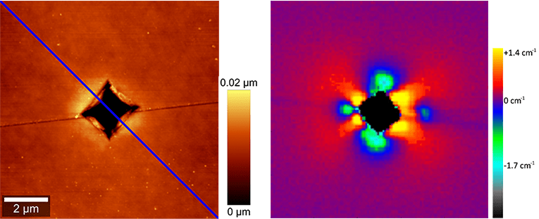
alpha300 RA Raman AFM stress in silicon
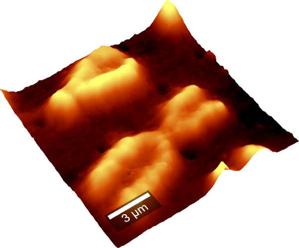
Correlative Raman-AFM microscopy of stress in silicon. Left: 10 μm x 10 μm AFM topography image and depth profile (bottom). Right: Corresponding Raman image revealing the stress areas in silicon.

Specifications

Raman General Operation Modes

  • Raman spectral imaging: acquisition of a complete Raman spectra at every image pixel
  • Planar (x-y-direction) and depth scans (z-direction) with manual sample positioning
  • Image stacks: 3D confocal Raman imaging
  • Time series
  • Single point Raman spectrum acquisition
  • Single-point depth profiling
  • Fibre-coupled UHTS spectrometer specifically designed for Raman microsopy and applications with low light intensities
  • Confocal Fluorescence Microscopy
  • Bright Field Microscopy


AFM General Operation Modes

  • Contact Mode
  • AC Mode (Intermittent Mode, Phase Imaging)
  • Digital Pulsed Force Mode (DPFM)
  • Lift Mode™
  • Magnetic Force Microscopy (MFM)
  • Electrostatic Force Microscopy (EFM)
  • Force Distance Curves
  • Nanomanipulation and Lithography
  • Lateral Force Microscopy (LFM)
  • Kelvin Probe Microscopy
  • Chemical Force Microscopy (CFM)
  • Current Sensing Mode
  • others on request


Computer Interface

  • WITec software for instrument and measurement control, data evaluation and processing

Basic Microscope Features

  • Research grade optical microscope with 6x objective turret
  • Video system: video CCD camera
  • High sensitivity b/w video camera to view sample and AFM tip in transmission
  • LED white-light source for Köhler illumination of AFM tip and sample
  • Manual sample positioning in x- and y-direction
  • Fibre coupling
  • Microscope base with active vibration isolation system


Raman Optional/Upgradable Operation modes

  • Additional lasers, several wavelenghts eligible
  • Additional UHTS spectrometers (UV, VIS, NIR)
  • Automated, motorized sample positioning and measuring with piezo-driven scan stages
  • Automated confocal Raman imaging
  • Automated multi-area and multi-point measurements
  • Ultrafast Raman imaging, optional available
  • Upgradable for epi-fluorescence applications
  • Adapter for higher samples
  • TrueSurface for Raman depth profiling
  • Autofocus


TERS

WITec’s unique alpha300 RA microscope systems with Raman and AFM techniques combined in one instrument provide optimized instrumentation for TERS experiments.

The WITec TERS advantages

  • Unique integrated Raman-AFM technique from one source for reliable TERS experiments
  • Flexible sample illumination from either above, below, or from the side

Contact

Please fill in all data fields to ensure we can process your inquiry as quickly as possible.


Related Applications

Food TechnologyForensicsArchaeology & The ArtsCoatings & Thin FilmsEnergy StorageGeo ScienceLife ScienceMaterials ScienceNano-Carbon & 2D MaterialsPharmaceutics & CosmeticsPolymersSemiconductors & Photovoltaics