Acquire research-grade results
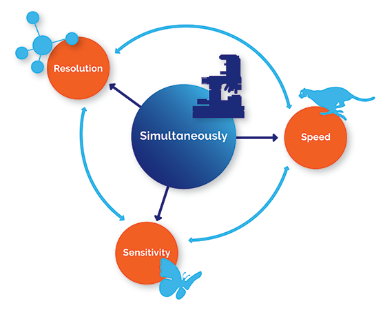
Optimised optics throughout the entire beam path deliver class-leading speed, sensitivity, and resolution for leading-edge research and development
The Oxford Instruments witec360 is the new benchmark for Raman imaging and correlative microscopy systems, designed to meet the growing demands of academic and industrial researchers for comprehensive analysis at the nanoscale.
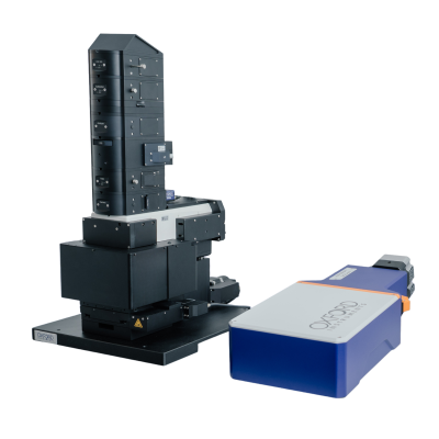
The witec360, successor to the pioneering alpha300 series, sets new standards in performance and broadband capability. Its fundamentally modular design provides immediate and long-term value with easy onboarding for Raman newcomers and the versatility to keep pace with varied and quickly evolving applications.
Acquire research-grade results
Optimised optics throughout the entire beam path deliver class-leading speed, sensitivity, and resolution for leading-edge research and development
Experience unprecedented versatility
Broadband capabilities provide the spectral flexibility needed for demanding research tasks and diverse applications in multi-user facilities
Gain comprehensive insight
Seamless integration of imaging techniques enables the correlation of chemical and structural properties for a more thorough understanding of samples
Configure your microscope for current and evolving experiments
Modular design offers a wide range of capabilities and upgrade options to deliver tailored solutions and scalability to match your institution’s requirements and resources
Enhance productivity with intelligent automation
Advanced hardware automation and intuitive software simplify data acquisition and analysis for operational efficiency and fast onboarding of users with different experience levels
Ensure consistency and transparency
Multi-user management, internal calibration, automated reporting, and streamlined analysis facilitate reproducibility and support institutional and regulatory compliance
Advanced confocal Raman technology for every task:
Learn more about the confocal Raman imaging technique
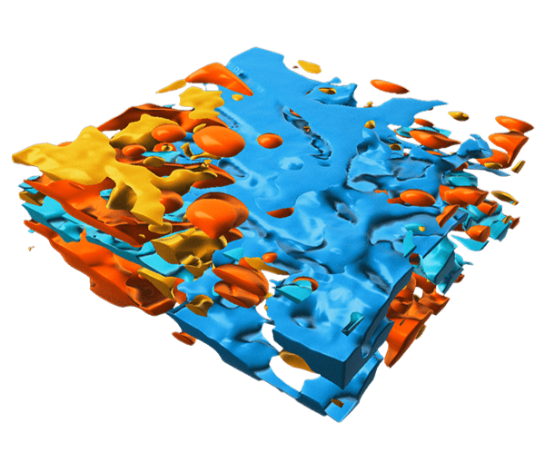
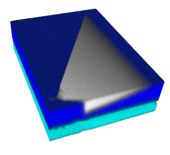
3D Raman image of a cosmetic emulsion rendered in Imaris. Dimensions: 30 x 30 x 10 µm³. Image credits: Oxford Instruments
Flexible – sustainable – future-proof
The witec360’s modular architecture empowers you to obtain the matching system for your specific experimental challenges. We provide solutions ranging from fixed configurations for single applications to specialised instruments for advanced academic research, as well as tools designed to meet industry standards and multi-user facility demands. Its flexible design ensures that the witec360 evolves and expands with your future requirements.
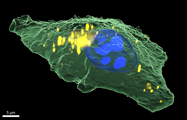
3D Raman image of a human epithelial cancer cell rendered in Imaris. Sample courtesy of Dr. Irina Estrela-Lopis and Tom Venus, Institute of Medical Physics and Biophysics, Leipzig University, Germany.
Choose your microscope basis (upright/inverted) |
Select your microscope components |
Incorporate advanced |
Customise with accessories |
By integrating correlative techniques, the witec360 expands its analytical capabilities and delivers complementary insights about chemical, topographic, mechanical, and structural properties at the same sample location. Its software ensures user-friendly control, effortless result correlation, and intuitive image overlay for streamlined analysis workflows.
Both witec360 systems and microscopes from the previous alpha300 series can be upgraded to include correlative imaging techniques.
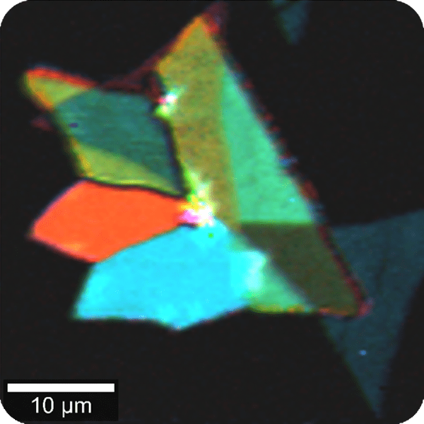
SHG is sensitive to non-centrosymmetric structures and is ideal for analysing crystal properties in 2D materials and label-free insights in life sciences.
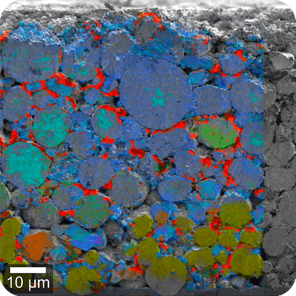
SEM techniques add nano-structural, crystallographic, and elemental information, especially valuable for material analysis, nanotechnology, and geosciences.
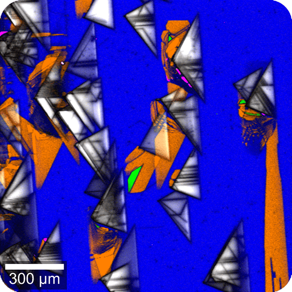
PL provides critical insights into optoelectronic properties, which makes it a widely used technique in material science and semiconductor characterisation.
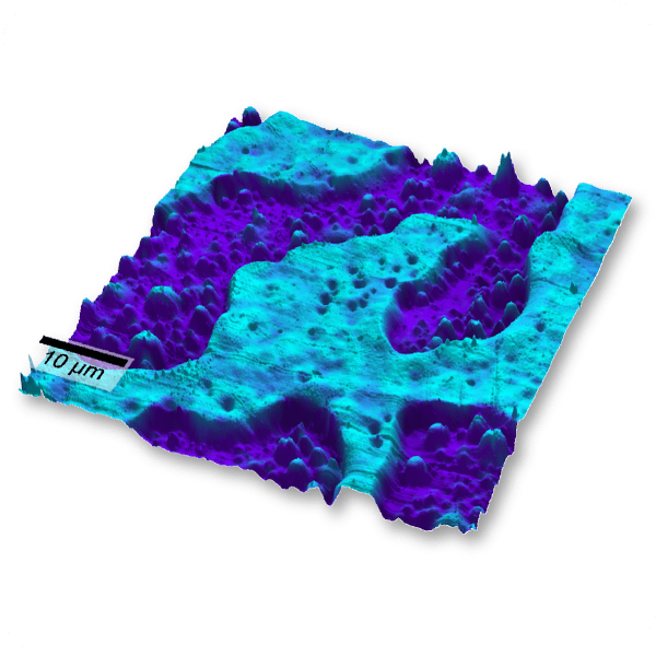
AFM reveals nanoscale surface properties, such as topography, adhesion, and stiffness, that are particularly important in materials science and nanotechnology.
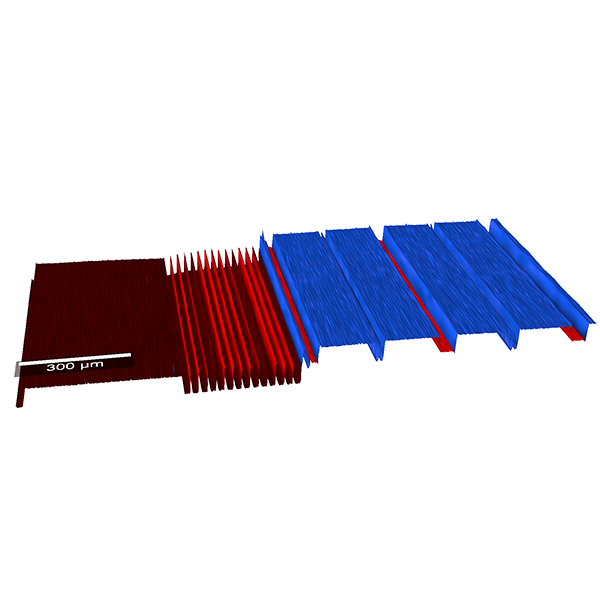
TrueSurface enables simultaneous topographic and Raman measurements of samples with irregular surfaces, such as rocks, tablets, or micro-structured materials.
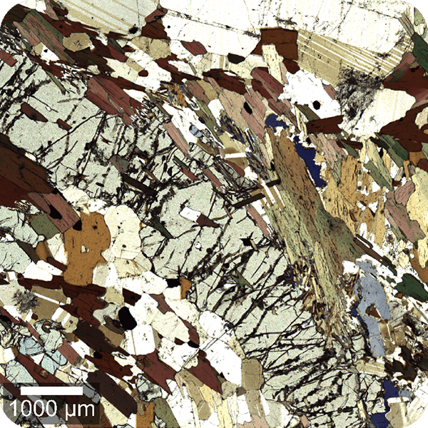
To efficiently locate and visualise regions of interest, witec360 microscopes feature Köhler illumination for class-leading white-light images in various modes.
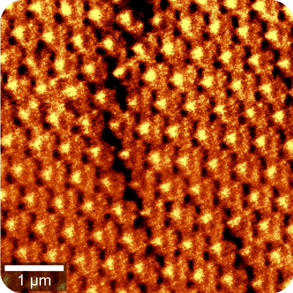
SNOM achieves optical imaging beyond the diffraction limit, with resolutions down to ~ 60 nm, suited for analysing nanomaterials and complex samples.
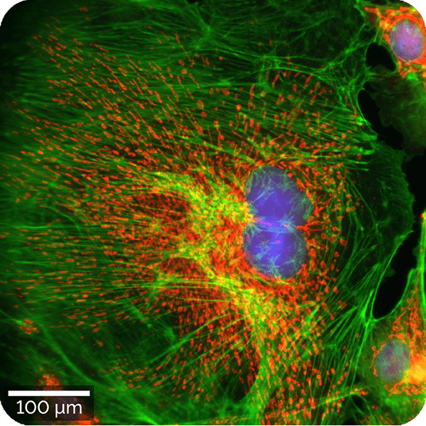
For specific applications in life sciences that rely on fluorescent labelling, Raman data can be complemented with fluorescence microscopy.
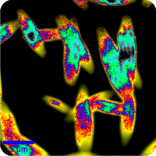
TCSPC techniques such TLM and FLIM provide valuable insights in semiconductor and optoelectronics research, as well as life sciences.
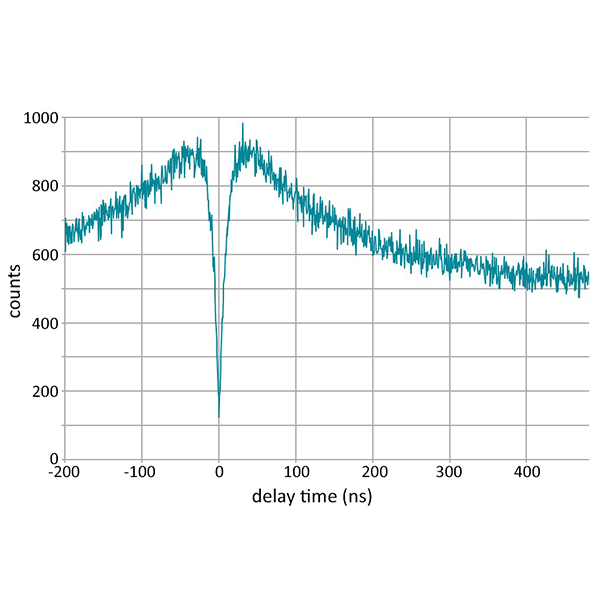
Correlative Raman, PL and antibunching enables efficient identification of single-photon emitters (SPEs) critical for quantum computing and cryptography.
The witec360 sets the highest standards for performance in confocal Raman imaging. Every component is optimised for maximum transmission efficiency and consistency, enabling accurate results within seconds. This accelerates your imaging workflows and facilitates even the most challenging applications.
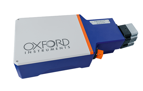
Hexalight - the core of witec360’s performance
Essential to the witec360 is the innovative Hexalight spectrometer, equipped with lens-based, mirror-free optics and 6 grating positions for optimal throughput and spectral flexibility across the broadest wavelength range.
Read more about Hexalight here.
Maximise efficiency and simplify your workflows with advanced automation options for witec360 microscopes that help ensure consistency and deliver high-quality results you can rely on.
Multi-User Labs: Accommodate varying experience levels and streamline workflows with easy-to-use shared equipment. |
Industry Researchers: Handle recurring experimental tasks quickly and precisely to accelerate development. |
Raman Newcomers: Simplify advanced tasks and enable confident operation with intuitive onboarding. |
Raman Experts: Push the boundaries with advanced automation for complex experiments. |
Feature |
Benefit |
AutoBeam – One click precision alignment Automatic laser beam path alignment using a certified reference sample, ensuring diffraction-limited resolution and stable performance. It features a memory function for multiple laser alignments and enables fully remote operation. |
Ideal for multi-user labs and enclosed setups, enabling hands-free, repeatable alignment for consistent, high-quality results. |
TrueCal – Calibration made easy Automated pre-configured calibration routines, keeping your system spectrally aligned and experiment ready. |
Perfect for repetitive or regulated workflows requiring precision, compliance, and quantitatively accurate results. |
Feature |
Benefit |
TrueComfort – Simplify everyday operations Enhanced convenience and accelerated imaging through streamlining essential operations:
|
Budget-conscious solution for increasing efficiency and ease-of use in routine imaging tasks. |
TrueSurface – Always in focus Actively maintains perfect focus during the entire measurement for sharp and high-quality images. |
Ideal for large-area scans and samples with curved, tilted and structured surfaces. |
Motorised laser coupler – Expand your wavelength options Software-controlled switching between up to six laser wavelengths (UV to NIR). |
Perfect for multi-wavelength experiments and diverse samples requiring varied excitation conditions. |
TruePower™ – Absolute laser power control Sets and controls absolute laser power within the measurement with an accuracy of <0.1 mW (down to 0.1 µW in extended configurations) for consistent, reproducible illumination. |
Essential for energy-dependent experiments and sensitive samples requiring strict laser power control. |
Motorised Polarisation Modules – Effortless and accurate Automated adjustment of freely rotatable excitation polarisers and detection analysers, streamlining polarisation-sensitive and polarisation-resolved series measurements. |
Ideal for molecular orientation or anisotropic materials studies, with improved usability and time efficiency. |
Motorised and piezo-driven stages – Precision made easy Automatic positioning of samples with motorised and piezo-driven scan stages. |
Essential for large area mapping, automatic zooming into regions of interest and revisiting of defined measurement positions. |
Our advanced software streamlines data processing and analysis, allowing you to quickly interpret results.
Feature |
Benefit |
Software Suite Analysis Wizard Simplifies data analysis with guided, click-through workflows. |
Reduced training time and accelerated data analysis for users of all experience levels. |
TrueComponent analysis Automatically identifies and differentiates sample components in an image. |
Fast and comprehensive analysis of complex samples, easy spectral unmixing. |
|
Quickly matches measured spectra to known molecules using renowned spectral databases. |
Direct peak assignment without the need of interpretation or mathematical evaluation. |
From large-area composition mapping to 3D stress and defect analysis, the witec360 drives material science innovations across polymers, geological samples, nanomaterials, chemistry and beyond. Its versatility supports advanced in-situ studies and correlative techniques, enabling comprehensive insights from material research to production.
The witec360 advances semiconductor innovation by integrating high-resolution Raman and photoluminescence imaging for detailed metrology covering structural and electronic insights. This includes non-destructive profiling, stress field mapping, defect characterisation and bandgap analysis.
The witec360 empowers life science research with label-free and non-destructive Raman imaging in 2D and 3D for detailed analysis of cellular structures and processes. It provides insights into metabolism, drug uptake, lipid and protein organisation, enabling comprehensive studies of biological interactions and disease mechanisms.
For pharmaceutical R&D, the witec360 Raman microscopes offer high-resolution insights into drug formulation, stability, and bioavailability. From API mapping and polymorph distribution in various dosage forms to surface coatings and uptake mechanisms, the witec360 ensures fast, precise results to accelerate time-to-market.
If you have any questions, or would like to speak to an expert or request pricing, we will reply to your enquiry shortly.
Get In Touch